
All
Products
Resources
News
FAQ
Search

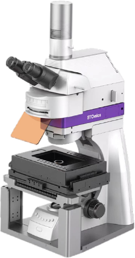


Go Optical | |
Optical System | Positive fluorescence microscope |
Objective Lens | 4X, 10X, 20X, (40X) universal motorized objective lens converter, support expansion |
Observation Methods | Epi-fluorescence, Epi-bright field, Transmitted bright field |
Travel Range X&Y | 100 mm x 70 mm |
Fluorescent Channel | Standard configuration: DAPI, FITC, TRITC, CY5, and supports optional configuration of other dye filter blocks |
Auto-focusing | Two optional modes——Auto-focus mode and map navigation mode |
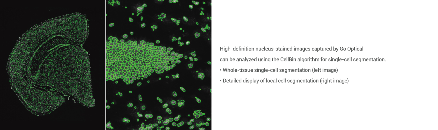
Working Efficiency | Scanning 10mm x 10 mm under 10X objective lens, scanning time≤70 s |
Working Mode | Manual operation by human eye observation; fully automatic scanning and imaging. |
Power & Dimensions & Weight | Working Environment |
Voltage: ~220 V Rated Power: 300 VA Dimensions (W x H x D): 400 mm (L) x 334 mm (W) x 726 mm (H) Weight: ~36 kg | Temperature: 5℃~40℃ Humidity: 30% RH to 75% RH, with no condensation |
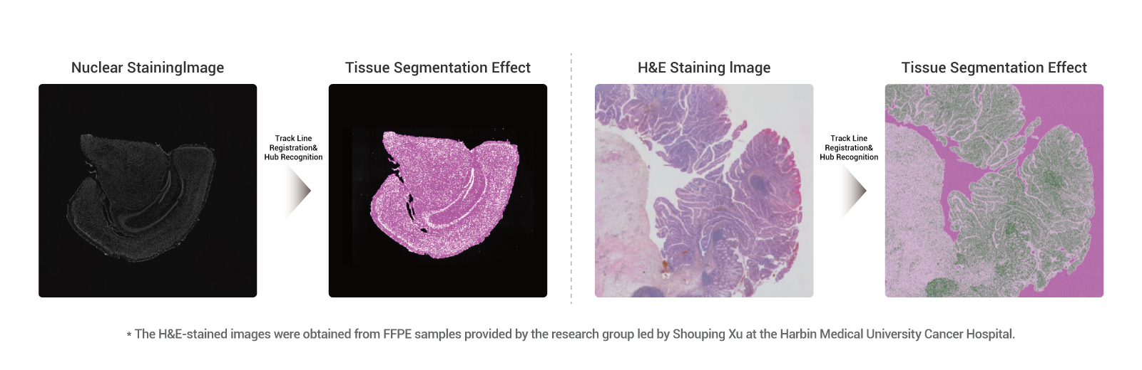
The Go Optical is primarily designed for various application scenarios within spatiotemporal omics, including:
1)Fluorescence imaging mode for capturing fluorescence-labeled silicon chip samples;
2)Brightfield imaging mode for capturing pathology-stained silicon chip samples;
3)Transmission brightfield imaging mode for capturing pathology-stained glass slide sample.
The Go Optical has currently obtained RUO-CE and RUO-NRTL certification, allowing to be sold in such as China, North America, the EU and Asia -Pacific.
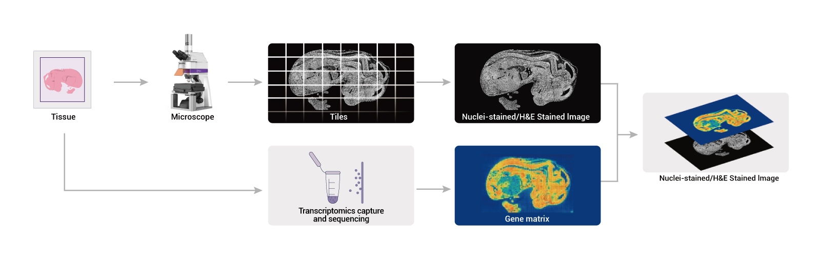
Go Optical incuding 2 parts, hardware and software. The hardward comprises a microscope, an electronic control box, a computer host, and a display monitor. 2 softwares are Scanncer and Tiff Browser.