The human brain has difficulty recovering on its own after an injury, but the brain of the Ambystoma mexicanum can. Brain regeneration is a complex biological process. Which kinds of key biological changes that occur during this process, and what are the important cells involved in this process, and their functions?
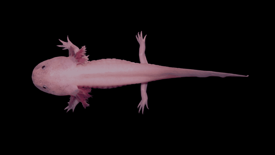
The research team led by the BGI Research in Hangzhou, China, together with scientists from 17 institutions in 3 countries, was used to analyze and compare the brain development and regeneration processes of Ambystoma mexicanum, and to construct the first spatial and temporal atlas of brain regeneration in Ambystoma mexicanum, which is also the first spatial and temporal atlas of brain regeneration in the world.
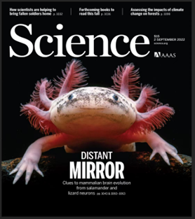
Through the study, the research team identified key neural stem cell subpopulations in anatomy brain regeneration process, depicted the process of such stem cell subpopulations to remodel damaged neurons, and also found that brain regeneration has some similarity with the developmental process, providing a boost to the cognitive brain structure and developmental process, and providing new directions for regenerative medicine research and treatment of the nervous system. In September 2(nd) 2022, the results were published as a cover article in the top international academic journalScience.

Science journal cover
So far, in just six months, the research results related to BGI Stereo-seq and single-cell technology have been published four times in Cell, Nature and Science.

Screenshot of Science official website
In order to understand how the brain regenerates, the team first had to understand how the brain develops. So, the team used Stereo-seq which is known as a super wide-angle 10-billion pixel "life camera," to "take pictures" of the anatomy's brain during six important periods of development. "This set of "photos" constitutes a spatio-temporal map of the anatomy's brain development. Through them, the team was able to obvious the molecular characteristics and spatial distribution dynamics of various types of neurons in the anatomy brain during the development process. It was found that the anatomy brain begins to specialize in spatially distinct neural stem cell subtypes from adolescence onward.

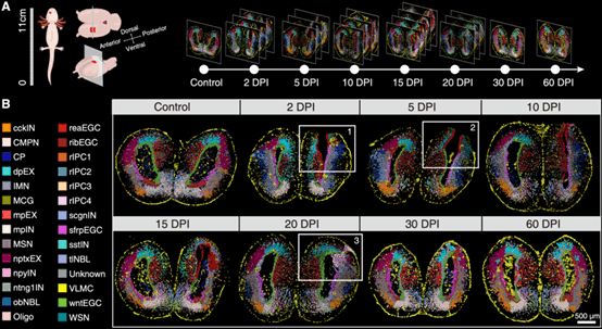
How does the process of regeneration after brain damage work? The research team performed mechanical damage surgery on the cortical region of the anatomy brain and "photographed" the brain samples by Stereo-seq at days 2, 5, 10, 15, 20, 30 and 60 after the damage which completely record the whole process from damage to regeneration and repair. By the way, the resulting film is much clearer than an X-ray, not only to see the shape of the brain, but also to continuously zoom in and see the cells in the brain, as well as the state of molecular changes in the cells.

By comparing regeneration "photos" over seven periods and the state of the wound during the process, the team found that a new subpopulation of neural stem cells emerged in the wound area early in the injury, and that this important group of cells was transformed by other neural stem cell subpopulations in the vicinity of the injury area after stimulation by the injury, and that during the subsequent regeneration process, new neurons were born to fill in the missing neurons at the injury site. In addition, although the wound was gradually filled with new tissue early in the repair process, it was not until day 60 after injury that the "photo" showed that the cell type and spatial distribution of the injured area returned to that of the uninjured side.

Spatio-temporal mapping of brain regeneration in Ambystoma mexicanum (image via Science)
Finally, the researchers also compared neuronal formation during brain development and regeneration in Ambystoma mexicanum and found that this process was highly similar during regeneration and development, and perhaps brain injury induced a reverse transformation of anatomy neural stem cells back to a developmentally rejuvenated state to initiate the regeneration process.
Dr. Ying Gu, co-corresponding author of the paper and a researcher at the BGI Research in Hangzhou, said, "The anatomy is evolutionarily more advanced compared to other Scleractinia fishes and has a higher similarity to mammalian brain structures. At the same time, its gene coding sequence is extremely similar to that of humans, and studying the initiation mechanism of brain regeneration in Ambystoma mexicanum and discovering the key genes involved may provide important guidance for the repair of neurological damage or degenerative diseases in humans."
The key genes in the regeneration process of the anatomy brain are said to be present in the human gene sequence as well. So why does it not play a regenerative role as it does in the anatomy brain? This may be a topic for scientists to study next.
Dr. Xiaoyu Wei, the first author of the paper and the researcher at the BGI Research in Hangzhou said that "This study was conducted primarily based on Stereo-seq, which achieves nanoscale subcellular resolution and combined with the large size of anatomy cells, which allows researchers to resolve important cell types in this process of anatomy brain regeneration at single-cell resolution and to track the spatial trajectory of their cellular lineage changes."
Xu Xun, co-corresponding author of the paper and director of the BGI Research said that "the construction of a spatio-temporal cellular atlas of anatomy brain development and regeneration is important for our understanding of brain regeneration as an important life process, amphibian brain structure, and the evolution of brain structure, and provides a new direction for our search for effective clinical treatments to promote self-repair and regeneration of human tissues and organs, as well as a valuable data resource for species evolution research ." And he also said that" we will also use Stereo-seq to explore the development and regeneration processes of more organs and species, and find the key regulatory mechanisms in the regeneration process to help the development of human regenerative medicine in the future,."


