
All
Products
Resources
News
FAQ
Search
01/08/2025
.jpg)
Spatial multi-omics technologies are transforming our understanding of complex biological systems with unprecedented detail. STOmics' Stereo-seq offers nanoscale resolution and an expansive field of view of up to 160 cm². In this issue, we highlight recent advances, key resources, and notable publications that showcase the power of Stereo-seq across neuroscience, disease research, and beyond.
With 212 publications to date, STOmics's Stereo-seq is empowering more researchers in developmental biology, neuroscience, disease research, and others.
Scientists created the world's first 3D digital mouse embryo at single-cell resolution, revealing critical signals in heart and foregut development—a leap forward in understanding birth defects. (Read in Cell)
A complete 3D single-cell atlas of fruit fly development, unlocking molecular insights for developmental biology & disease research. (Read in Cell )
Discovery of the "missing retinoic-acid switch", enabling enhanced tissue regeneration—a potential game-changer for regenerative medicine! (Read in Science)

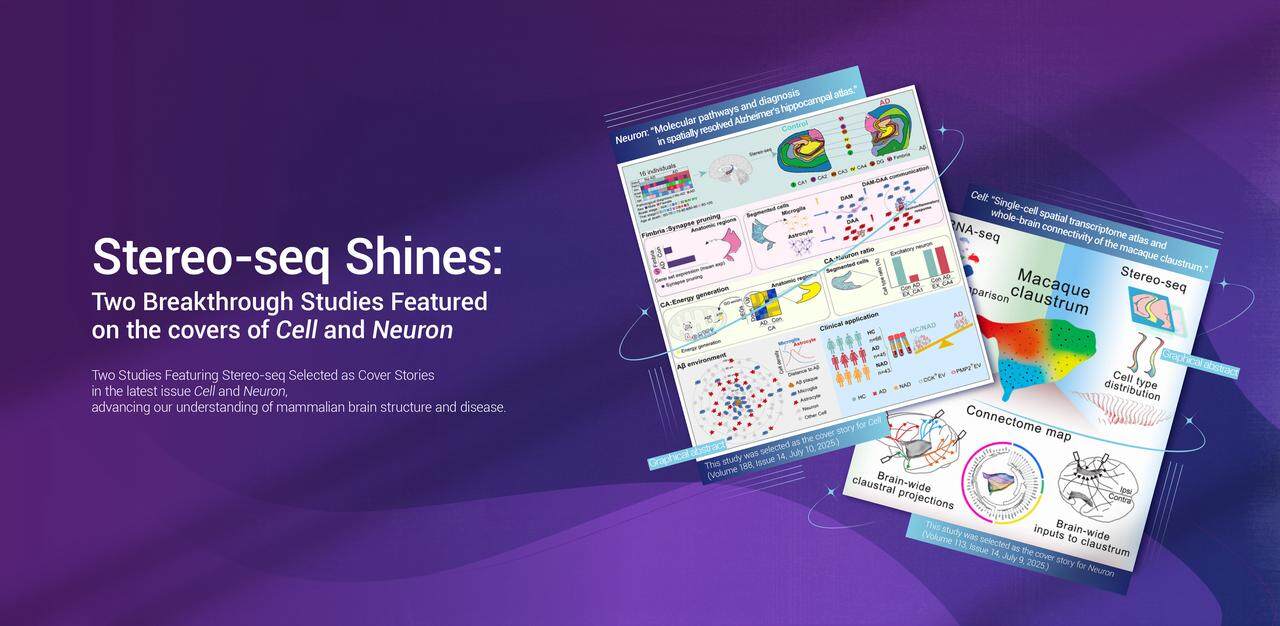
Two landmark studies leveraging STOmics' Stereo-seq spatial transcriptomics technology earned cover features in Cell and Neuron, showcasing its transformative impact on brain research.
Key Highlights:
Macaque Claustrum Study (Cell Cover) – The first panoramic molecular and circuit map of a brain region linked to consciousness and cognition, offering new insights into brain-wide information processing. (Read in Cell)
Alzheimer's Disease Decoding (Neuron Cover) – Spatially resolved molecular signatures reveal novel disease mechanisms, opening doors for diagnostics and therapies. (Read in Neuron)
These studies are part of ten high-impact publications from the Mesoscopic Brain Mapping Consortium, published across Cell, Neuron, and Developmental Cell, further validating Stereo-seq's precision and scalability in neuroscience.
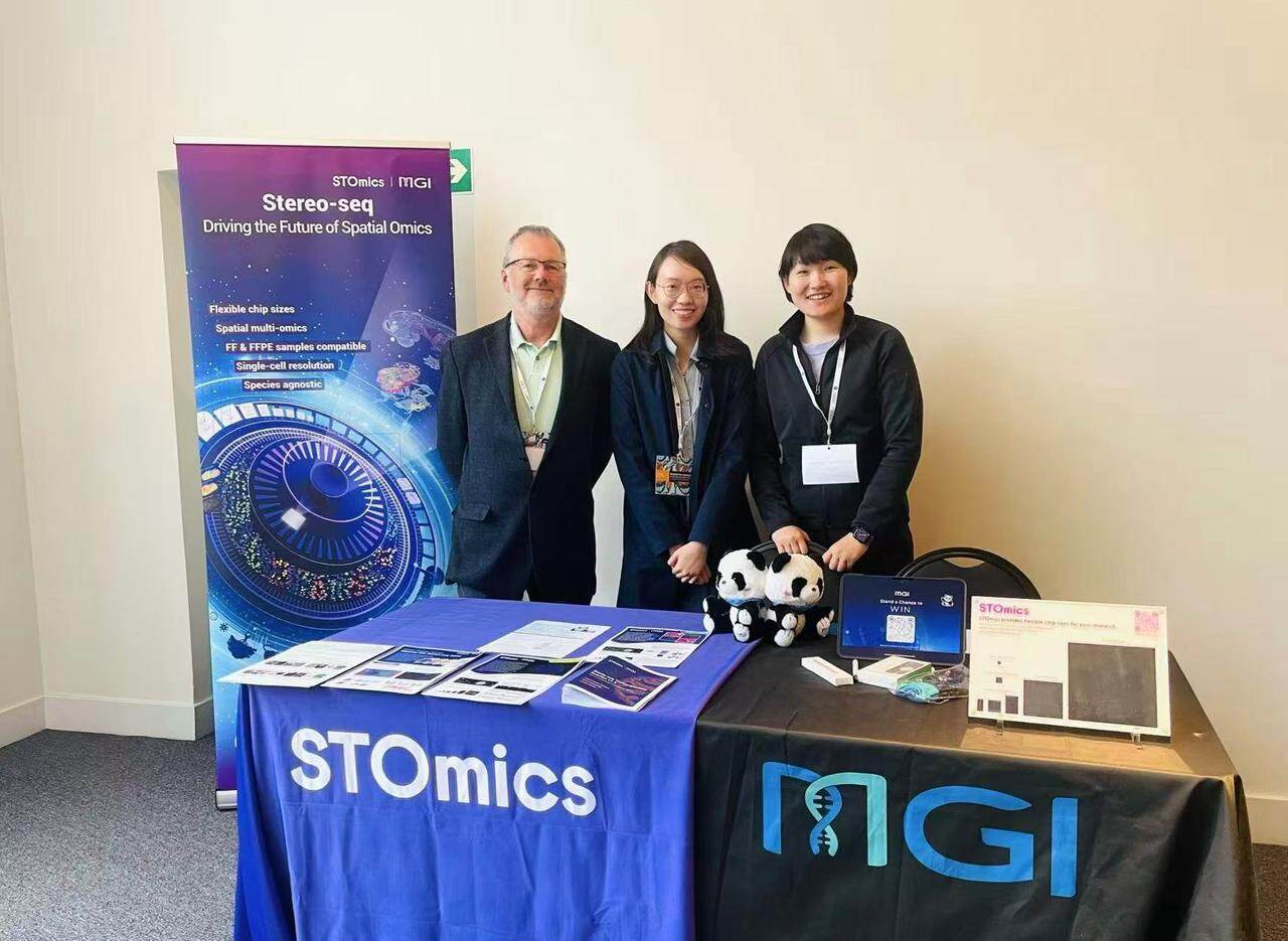
ESHG2025 in Milan, held from May 24–27, 2025, it was an inspiring event where together with our distributor MGI, we showcased Stereo-seq.
Highlights included Dr. Li Jiankang's presentation on "Unveiling spatial heterogeneity in medulloblastoma" , and Dr. Ji Yi's e-poster on "Analysis of Macrophage Spatial Distribution and Tumor Microenvironment in Esophageal Squamous Cell Carcinoma Using Stereo-ClTE-seq". It was a great platform for sharing advancements in spatial omics and connecting with industry leaders. We thank everyone who visited and look forward to future collaborations in this exciting field!

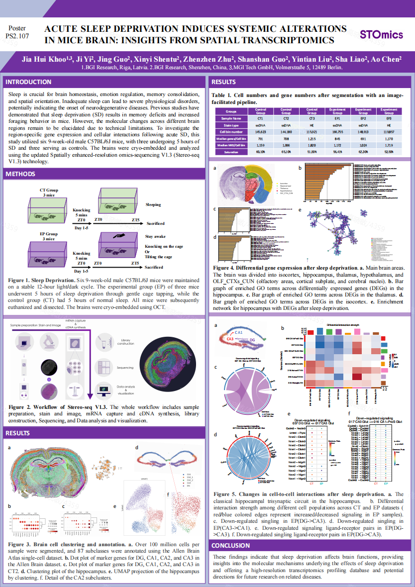
NeuroFrance 2025 in Montpellier, held from May 14–16, 2025, it was an exciting event where we showcased the latest advancements in Stereo-seq spatial transcriptomics technology. We also featured a poster on "Acute Sleep Deprivation Induces Systemic Alterations in Mice Brain," demonstrating how Stereo-seq V1.3 helps uncover gene expression changes and cellular interactions across brain regions—a key step toward understanding sleep's impact on brain function.
In June 2025, Edinburgh hosted the innovative EMBO Workshop focused on advancing spatial omics technologies. Researchers gathered to explore unprecedented resolutions in spatial biology, with highlights including the Stereo-seq talk titled “Precision Mapping with STOmics Stereo-seq: Whole-Transcriptome Spatial Profiling Across Species and Scales.”
In May, STOmics proudly announced ProfileXpert as its first CSP partner in France, marking a significant milestone in expanding access to Stereo-seq spatial transcriptomics across Europe. As part of our growing CSP program, this alliance exemplifies our commitment to connecting global partners with cutting-edge spatial omics solutions. We welcome more partners to join our CSP program.
.JPEG)
No worries if you missed our "Precision in Space: Stereo-seq Learning Series webinar" and "Stereo-seq Customer Sharing Series webinar" - the recordings are now available for immediate viewing.
Stereo-seq delivers spatially resolved ultra-high-resolution whole transcriptome datasets, so annotation algorithms may not scale well with increasing data size. The complexity of spatial context also hinders researchers from doing annotation effectively. In this webinar, Dr. Zheng Zhong will break down computational strategies to tackle this.

Spatially resolved transcriptomics unlocks the secrets of how cells interact with their environment through gene regulation. By analyzing spatial gene co-expression, we can uncover co-regulatory patterns and their biological significance.

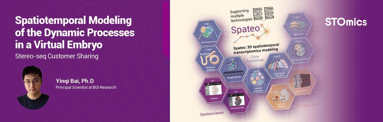
Embryogenesis holds the key to congenital diseases—but how do we quantify its spatiotemporal dynamics? In this webinar, we'll showcase Spateo - a revolutionary 3D modeling framework.
Unlock the full potential of your Stereo-seq data with a deep dive into essential image processing. While the SAW pipeline automates analysis, understanding each step is key for accurate interpretation and effective troubleshooting. Learn the fundamentals behind these critical stages, empowering you to confidently navigate your spatial transcriptomics results.
Streamline your Stereo-seq workflow and conquer common analysis challenges! Building on essential image processing steps, discover practical solutions for issues like failed Image QC, registration errors, and segmentation problems. We'll show you how to leverage StereoMap's powerful tools and third-party integrations to get your spatial transcriptomics analysis back on track.
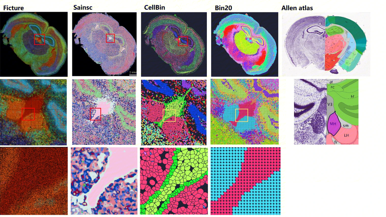
To empower your research, we've benchmarked eight popular community cell segmentation tools against our in-house method. Our comprehensive study, leveraging the CellBinDB dataset and tested across DAPI, ssDNA, H&E, and mIF staining, provides data-driven recommendations to help you select the optimal algorithm for your specific experimental needs.
Ensure FFPE (Formalin-Fixed Paraffin-Embedded) section preparation and safe shipping with these expert guidelines. Learn key steps for optimal sample selection, sectioning, and transport to preserve tissue integrity for research.
Ensure H&E staining and proper evaluation of FFPE (Formalin-Fixed Paraffin-Embedded) sections with these expert guidelines. Learn key steps for selecting optimal tissue sections, assessing staining quality, and troubleshooting common issues.