
In the rapidly advancing field of spatial biology, context is everything. The ability to map gene expression within its native tissue architecture has transformed our understanding of cellular heterogeneity. However, for years, researchers have been confined by a physical boundary: the "standard" capture area. Most spatial transcriptomics platforms are limited to the small window. While this is sufficient for small biopsies or specific sub-regions, it often forces a compromise when studying larger biological systems.
When someone cut a tissue to fit a small chip, they lose the "neighborhood." They miss the transition zones between a tumor and healthy tissue, the systemic connectivity between organs in a developing embryo, and the intricate anatomical continuity of a whole brain. So, STOmics has introduced the Stereo-seq Transcriptomics Solution for Large Chip Designs (LCD) to bridge this gap. This solution scales up the field-of-view (FOV) to a macro scale without sacrificing the nanoscale resolution that Stereo-seq is known for.
What Large Chip Designs Actually Offer?
Stereo-seq LCD is the first spatial transcriptomics solution to combine a centimeter-scale field of view with true nanoscale resolution—a distinction that matters enormously in practice. The platform currently offers three standardized chip sizes: 1 cm × 2 cm, 2 cm × 2 cm, and 2 cm × 3 cm. For projects with exceptional scale requirements, customized configurations are available up to 13 × 13 cm, subject to project design and regional support agreements.
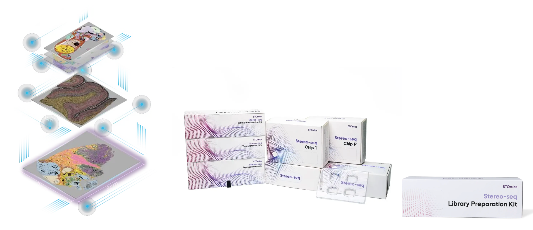
Figure 1. Stereo-seq Large Chip Designs (LCD)
Underlying all chip sizes is the same DNA Nanoball (DNB) patterned array technology: spatially barcoded probes with a spot size of 220 nm, center-to-center spacing of 500 nm, and unbiased capture of the poly-adenylated transcriptome in situ. There is no trade-off between area and resolution. The 2 × 3 cm chip captures six times more tissue than the standard 1 × 1 cm format while maintaining single-cell and subcellular sensitivity across the entire surface.
All three chip sizes are compatible with fresh frozen (FF) samples from any species. The compatible kit—the Stereo-seq Transcriptomics Set V1.3 for Large Chip Designs—supports both H&E and ssDNA staining workflows within the same protocol, and the total bench time from cryosection to sequencing-ready library runs approximately 1.5 days. Beyond whole-organ profiling, the larger capture area also enables running multiple co-embedded tissue sections in parallel on a single chip, serial section capture for 3D transcriptome reconstruction, and the spatial extent needed to resolve heterogeneity across functionally distinct tissue zones—cortical layers, tumor margins, skeletal compartments—all within a single experiment.
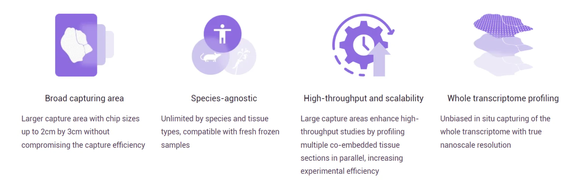
Figure 2. The Key Highlight of Stereo-seq LCD Solution
Use Case 1: Whole-Embryo Atlas at E18.5
On a 1 × 2 cm chip, a single sagittal section of a female C57BL/6 mouse embryo at gestational day E18.5 was profiled in its entirety. The section was stained with H&E on-chip, imaged, and processed through SAW v8.1 and StereoMap v4.1. At the Bin200 level, the median MID count per unit was 82,562, with a median of 10,952 gene types detected. At single-cell resolution (Cell Bin), the median gene count per cell reached 433 with 569 MIDs per cell—sufficient sensitivity to distinguish closely related progenitor populations.
Critically, the spatial context is preserved at every scale. Using the lasso tool in stereopy, individual organs within the embryo section—brain, liver, kidney, lung, inner ear, submandibular gland, gastrointestinal tract—were re-clustered independently, each generating refined cell-type maps with hundreds of gene types detected per cell. This multi-organ, single-section approach is only possible because the chip is large enough to hold the whole embryo. No stitching artifacts, no missing regions, no ambiguity about organ boundaries.
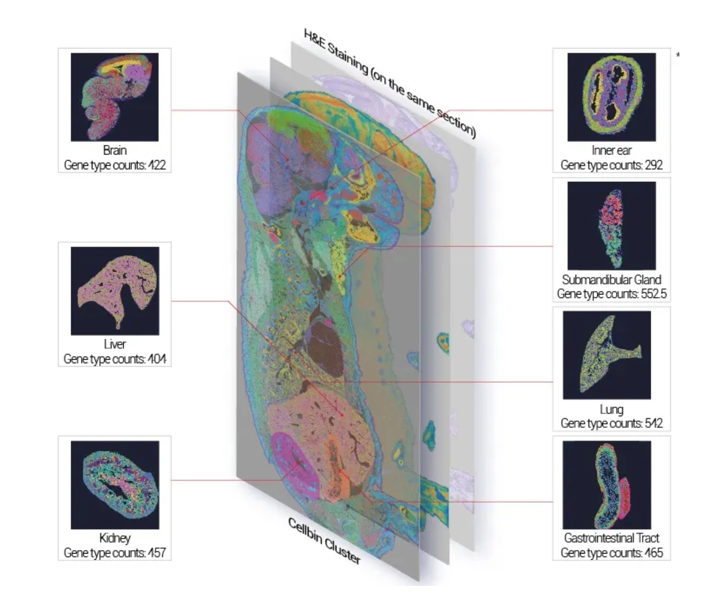
Figure 3. Spatial Gene Expression of Mouse Embryo Fresh Frozen Tissue. Stereo-seq LCD Solution enables the simultaneous detection of multiple organs within a single section, as well as precisely identifying different cell types within individual organ.
Use Case 2—Whole Rat Brain at Subcellular Resolution
A full coronal section of an adult Sprague-Dawley rat brain was profiled on a 2 × 3 cm chip, capturing both hemispheresand the major anatomical divisions of the CNS simultaneously. The tissue was stained with DAPI, and the dataset was analyzed with SAW v7.0 and StereoMap v3.0. The sequencing depth was substantial: 28 billion total reads, with a sequencing saturation of 93.6%—indicating deep, near-complete transcriptome coverage.
At CellBin resolution, the median gene count per cell was 514 with 1,031 MIDs per cell—sufficient to resolve fine-grained neuronal subtypes, glial populations, and vascular cell identities across the entire brain section. The fraction of MIDs falling under tissue reached 84.6%, confirming efficient, low-background capture across the full 6 cm² surface. This dataset demonstrates that Stereo-seq LCD delivers high-quality, biologically interpretable data even at the extremes of its format.
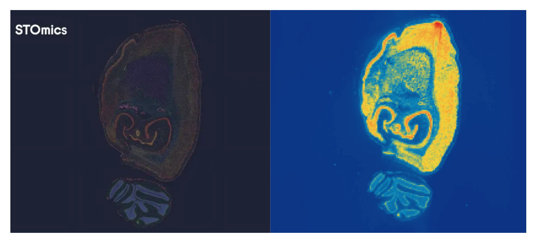
Figure 4. Spatial Gene Expression of Rat Brain Fresh Frozen Tissue. Stereo-seq LCD enables near whole-brain profiling at subcellular resolution.
Practical Notes: Preparing Large Tissues for LCD
Sample quality is the single most important variable in a spatial transcriptomics experiment, and the requirements are slightly more stringent for LCD than for the standard Stereo-seq workflow. A minimum RNA Integrity Number (RIN) ≥ 6.0 should be applied for LCD—higher than the RIN ≥ 4 threshold for standard V1.3 kits. Drawing from the Sample Preparation Guide for Fresh Frozen Samples, here are some further critical considerations for LCD experiments:
Specialized embedding molds: Large tissues require precise embedding to prevent curling or uneven freezing. Use stainless-steel base molds (mold A and B) specifically designed for 1×2, 2×2, or 2×3 cm formats.
Preventing deformation: A common challenge with large sections is compression during the freezing process. STOmics recommends using a pre-cooled steel ruler placed on the high side of the mold to act as a leveler, ensuring the tissue stays flat and undeformed as the OCT solidifies.
Eliminating air bubbles: Because the surface area is larger, the risk of trapped air bubbles increases. Always use a syringe to carefully remove any bubbles from the OCT before freezing, as bubbles can cause "holes" in your spatial data.
Optimal sectioning: Aim for a 10µm thickness. For LCD chips, maintaining a consistent temperature in the cryostat is vital to prevent the larger sections from shattering or "chattering" during the stroke.
Storage: Large tissue blocks are more susceptible to dehydration. Wrap them tightly in aluminum foil and store them at -80°C in a sealed plastic bag.
Standard vs. Large Chip: A Comparative Overview
Table 1. The Comparison between Standard Stereo-seq and Stereo-seq LCD
Ready to Scale Up Your Science?
Whether you are building a whole-embryo cell atlas, mapping the full spatial extent of a brain hemisphere, profiling multi-organ systems in a single section, or capturing large tumor microenvironments that exceed the boundaries of standard chips, Stereo-seq Large Chip Designs delivers the field of view your biology demands—without compromising the nanoscale resolution, transcriptome depth, or data quality that Stereo-seq is known for.
Everything you need to run Stereo-seq Large Chip Designs in your own laboratory is available as a complete kit solution. The full workflow—from permeabilization optimization through library preparation—is supported by the following reagent sets:
Table 2. Stereo-seq Kit Solution List for Large Chip Designs
→ Explore Stereo-seq Large Chip Designs and Download the Product Note PDF: Transcriptomics Large Chip Design - STOmics
→ Download the User Manual PDF: Stereo-seq Transcriptomics Set V1.3 for Large Chip Designs User Manual
→ Explore Demo Data: Mouse Embryo (E18.5, 1×2 cm) | Rat Brain (2×3 cm)
To learn more about how our spatiotemporal omics solutions can support your research, please feel free to Contact Us or reach out via email at info_global@stomics.tech.
FAQs
Q: Can I use Large Chip Designs with FFPE samples, or only fresh frozen tissue?
A: The current Stereo-seq Transcriptomics Set V1.3 for Large Chip Designs is validated for fresh frozen (FF) samples only. FFPE compatibility is not supported at this time. For best results, ensure your FF tissue has a RIN value ≥ 6.0 before proceeding with the LCD workflow.
Q: My tissue is larger than the 2×3 cm chip. Is there a bigger option?
A: Yes. While the three standard sizes (1×2, 2×2, and 2×3 cm) cover the majority of research applications, STOmics offers customized chip sizes up to 13×13 cm for projects with exceptional requirements. Availability depends on project design, laboratory equipment, and regional support agreements—contact your local STOmics representative or reach out at info_global@stomics.tech to discuss your specific needs.
Q: How does the data analysis workflow for LCD differ from standard Stereo-seq?
A: It doesn't—the downstream analysis pipeline is identical. Sequencing data is processed through the SAW (Stereo-seq Analysis Workflow) pipeline and visualized in StereoMap, exactly as with standard chips. The only practical difference is file size: larger capture areas generate proportionally larger datasets, so adequate computational storage and memory should be planned accordingly.