
The thymus is a small, bilobed organ nestled behind the sternum, yet it fundamentally dictates the balance of human immunity. As the essential training ground for the adaptive immune system, it is the sole reason your T cells target foreign pathogens rather than your own tissues. Every single T cell defending your body today was rigorously tested and certified within this transient organ (one that naturally shrinks after puberty). For decades, immunologists could profile the cellular composition of the thymus using traditional sequencing tools, but they were blind to a critical variable: the spatial coordinates of where these cells resided, their dynamic cell-cell interactions, and how this complex cellular geography shifts from fetal development into childhood. Today, next-generation spatiotemporal omics is changing that. By bridging the gap between high-resolution sequencing and physical tissue architecture, we are finally uncovering the precise spatial mechanisms underlying T cell maturation and revealing the structural secrets that shape central immune tolerance.
The Cortico-Medullary Axis and T Cell Maturation
Thymopoiesis is not a static event; it is a highly coordinated spatial migration driven by precise chemokine gradients. The functional architecture of the thymus is strictly segregated into two primary compartments: the outer cortex and the inner medulla.
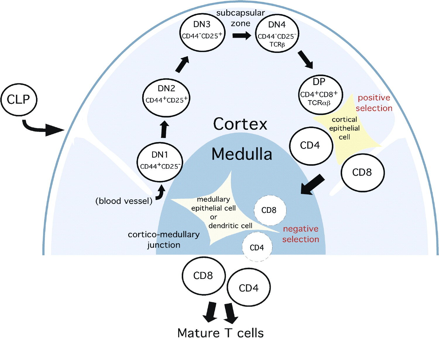
Figure 1. Migration of maturing T cells in the thymus (Lehar & Bevan, 2002)
Progenitor cells arrive at the cortex, where they undergo TCR gene rearrangement and enter the stage of positive selection. Here, cortical thymic epithelial cells (cTECs) present self-peptides bound to Major Histocompatibility Complex (MHC) molecules. Thymocytes that demonstrate functional recognition of self-MHC receive survival signals, while those that fail undergo death by neglect (Gamble et al., 2025).
Positively selected thymocytes then upregulate specific chemokine receptors (such as CCR7) and migrate toward the medulla for negative selection. In the medullary microenvironment, medullary thymic epithelial cells (mTECs) and dendritic cells present a vast array of self-antigens. Thymocytes exhibiting strong autoreactivity to these antigens are either eliminated via apoptosis (clonal deletion) or diverted into the regulatory T cell (Treg) lineage. This spatial transit ensures that only MHC-restricted, self-tolerant mature T cells are exported to the periphery.
Thymic Mimetic Cells: Expanding the Scope of Antigen Presentation
A central question in immunology has been how the medullary microenvironment can present antigens specific to spatially distant tissues—such as pancreatic islets, retinal proteins, or intestinal mucosa—to enforce comprehensive self-tolerance.
Historically, this ectopic expression of peripheral tissue antigens (PTAs) was primarily attributed to the Autoimmune Regulator (Aire) transcription factor. However, recent single-cell RNA sequencing (scRNA-seq) studies have identified a complementary, sophisticated mechanism within the post-Aire compartment: thymic mimetic cells (Givony et al., 2023).
Morphological observations of "misplaced" peripheral-like cells in the thymus date back over a century, often dismissed as developmental anomalies. Modern transcriptomics has redefined them as specialized mTEC subpopulations that adopt the transcriptional profiles and chromatin landscapes of diverse extra-thymic tissues.
Driven by lineage-defining transcription factors, these mimetic cells accurately reflect the phenotypes of specific peripheral tissues. For instance:
Tuft mimetic cells rely on Pou2f3 to mirror intestinal chemosensory cells.
Muscle mimetic cells upregulate terminal differentiation markers like Myog.
Neuroendocrine mimetic cells express Insm1, FoxA and Ascl1, mirroring sensory and neural tissue profiles.
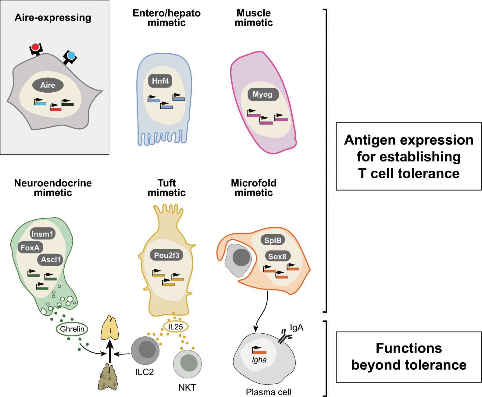
Figure 2. Mimetic cell characteristics and functions. Mimetic cells establish central T cell tolerance by deleting self-reactive T cells or promoting Treg differentiation. They also mimic extra-thymic roles: tuft mimetic cells secrete IL25, activating ILC2s to drive thymic regeneration; neuroendocrine mimetic cells produce ghrelin to regulate thymic cellularity; microfold mimetic cells support IgA⁺ plasma cell induction. These functions depend on lineage-specific TFs: Hnf4 (enterocyte-hepatocyte), Pou2f3 (tuft), Insm1 (neuroendocrine), and SpiB/Sox8 (microfold)(Huisman et al., 2025).
By generating these localized, highly specific transcriptional mimics within the medulla, the thymus ensures that developing thymocytes encounter a comprehensive representation of the body's cellular diversity, thereby suppressing autoimmunity before it can initiate.
The Limitations of Dissociation-Based Sequencing
While scRNA-seq was instrumental in the molecular identification of mimetic cells, it is inherently limited by the need for tissue dissociation. Enzymatic digestion destroys the spatial coordinates of the tissue, resulting in a critical loss of topological data (Li et al., 2024).
Through standard scRNA-seq, researchers can quantify the expression levels of mimetic cells, but fundamental questions remain unanswered:
What is the precise physical distance between specific mimetic cell subtypes and medullary landmarks like Hassall's corpuscles?
Which specific subsets of developing thymocytes colocalize and interact with muscle or neuroendocrine mimetic cells?
How do these spatial niches reorganize dynamically across developmental stages?
To decode the functional mechanics of central tolerance, researchers require a spatial methodology capable of profiling the entire organ without sacrificing single-cell or subcellular resolution.
Redefining Spatial Biology: The Stereo-seq Advantage
STOmics' Stereo-seq provides a transformative solution to the limitations of dissociation-based sequencing, offering unprecedented resolution and scale for mapping complex microenvironments like the thymus (Yayon et al., 2024).
Nanoscale Resolution: Utilizing DNA nanoball (DNB) technology with a capture spot size of 500 nm, Stereo-seq achieves genuine subcellular and single-cell resolution. This nanoscale precision directly answers the question of exact physical distances between rare mimetic cell subtypes and medullary landmarks like Hassall’s corpuscles, mapping molecular gradients that traditional micron-scale platforms inherently blur.
Centimeter-Scale Field of View: Thymic structures and cortical-medullary ratios change fundamentally over time. Stereo-seq’s large chip designs (up to 13 × 13 cm) allow for the capture of macro-scale, intact human tissue sections. This capacity makes it possible to array multiple samples or developmental timepoints on a single slide, eliminating batch effects and directly revealing how these complex spatial niches reorganize dynamically across developmental stages.
Multi-Omic Integration via Stereo-CITE: To determine which specific subsets of developing thymocytes interact with muscle or neuroendocrine mimetic cells, researchers must capture both cellular lineage and functional phenotypes simultaneously. The Stereo-CITE protocol enables the simultaneous capture of spatially resolved whole-transcriptome data and multiplexed expression of dozens of surface or intracellular proteins on the exact same tissue section, providing the multi-dimensional resolution needed to validate true receptor-ligand engagement at the single-cell interface.
Broad Sample Compatibility: Tracking the spatiotemporal evolution of the thymus from fetal development to childhood requires a platform that can accommodate diverse clinical specimens. Stereo-seq offers unmatched flexibility, from fresh frozen (FF) sections for high-yield discovery to the Stereo-seq OMNI for FFPE solution—which recovers highly sensitive spatial data even from low RNA Integrity Number (RIN), highly degraded archival pathology blocks, ensuring that precious, historically inaccessible cohorts can be fully charted.
STOmics' Stereo-seq Portfolio for High-Resolution Mapping
Studies of central immune tolerance in the human thymus—such as the precise geopositioning of lineage transcription factors within rare mimetic epithelial cells—highlight the critical need for technologies that can resolve complex cellular microenvironments within an intact tissue context. To address this, STOmics offers a comprehensive portfolio of spatiotemporal omics solutions powered by the Stereo-seq platform. Enabling transcriptomic mapping at 500 nm resolution across centimeter-scale tissue regions, and integrating protein analysis via Stereo-CITE, this platform empowers researchers to conduct precise analyses of immune organogenesis, cell-cell interactions, and dynamic tissue architecture.
Table 1. STOmics product matrix
Mapping the Human Thymus: Fetal Development to Childhood
Leveraging the combined capabilities of Stereo-seq and Stereo-CITE, a research team from the A*STAR Institute of Molecular and Cell Biology (IMCB) in Singapore has successfully constructed the first high-resolution spatial multi-omics atlas of the human thymus.
By profiling tissue across eight distinct developmental timepoints—from early fetal stages through pediatric development—the study delineates the exact spatial geopositioning of lineage-defining transcription factors within rare thymic mimetic cells. This research provides a definitive spatial framework for understanding how the thymic microenvironment orchestrates immune tolerance and how perturbations in this architecture may predispose individuals to autoimmune diseases.
Be There for the Data Debut
This article outlines the science. The webinar is where you see it firsthand.
On May 28, lead researcher Dr. Uma Kamaraj from A*STAR's Institute of Molecular and Cell Biology (Singapore) will present the full spatial atlas of the human thymus — walking through how Stereo-seq and Stereo-CITE were used to geoposition lineage transcription factors within rare mimetic epithelial cells across eight developmental timepoints. This is the first public presentation of this dataset.
📅 Date: Thursday, May 28, 2026
⏰ Time: 10:00–11:00 AM SGT (03:00 AM CEST | 11:00 AM JST/KST | 12:00 PM AEST)
💻 Format: Zoom Webinar — ~40 min talk + live Q&A 📖 Published in: Nature Communications
References